
Parts of a Tooth: Anatomy, Layers and Typical Sizes Explained
Enamel, dentin, pulp and cementum each do a different job. Here is how a tooth is built, what holds it in place, and how big front teeth usually are...
By Dr. Boris Zusin · Published October 23, 2026
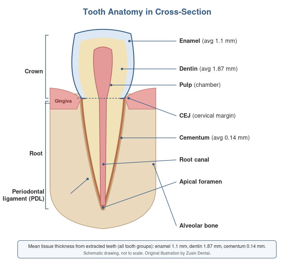
Every tooth looks simple from the outside, but each one is built from four different tissues, held in place by a support system of gum, ligament and bone. Knowing what's what makes it easier to understand why a cavity can be painless at first, why exposed roots feel sensitive, and why some treatments — like a root canal or a crown — are done the way they are.
The Four Tissues of a Tooth
Enamel
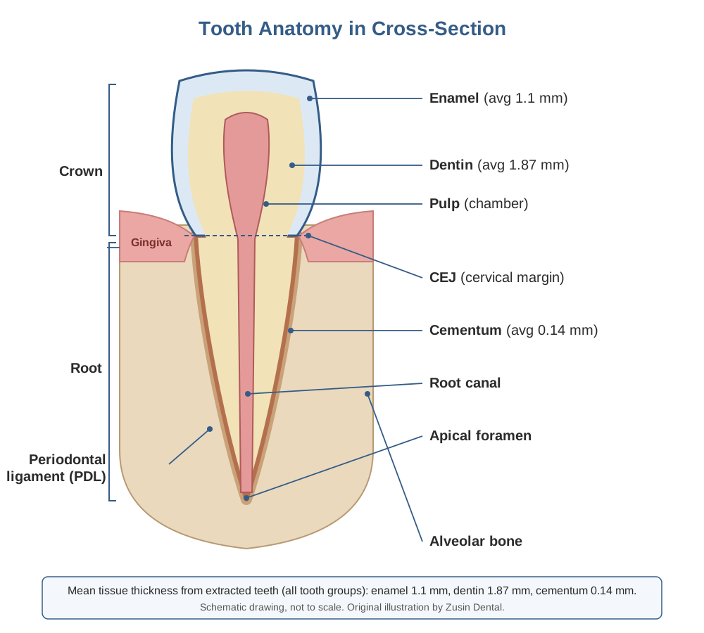
Enamel is the outer shell of the crown and the hardest tissue in your body. It is mostly mineral, which is why it is so strong and also why it cannot regrow once it is lost to decay or wear. Fluoride and a good home routine help harden and repair the surface, but a real hole needs a filling. In extracted teeth, enamel averages roughly 1.1 mm thick, and it is thickest on the molars.
Dentin
Dentin sits under the enamel and makes up most of the tooth, in both the crown and the root. It is softer than enamel and is threaded with microscopic channels that connect to the nerve. That is why sensitivity to cold or sweets starts when dentin is exposed by wear, a crack or receding gums. Dentin averages about 1.9 mm thick in extracted teeth. See All About Tooth Sensitivity for what to do about it.
Pulp
The pulp is the living center of the tooth: soft tissue with blood vessels and nerves, running from a chamber in the crown down narrow canals in the root. It exits at the tip of the root (the apical foramen), where it connects to the tissues around the tooth. When decay or a crack reaches the pulp, the result can be a toothache or an abscess, and a root canal is how the infected pulp is cleaned out while the tooth is kept.
Cementum
Cementum is a thin layer, about 0.14 mm on average, that covers the root and gives the fibers of the periodontal ligament something to attach to. It is much softer than enamel, so when gums recede and the root is exposed, it wears and decays more easily.
What Holds the Tooth In Place
The border between crown and root is the cementoenamel junction (CEJ), and healthy gum normally covers it. Below the gum line, the tooth sits in a socket of jawbone, cushioned by the periodontal ligament, a thin elastic layer that lets teeth move very slightly when you chew. Gum, ligament, cementum and bone together are called the periodontium, and gum disease is the process that breaks it down. This is why a tooth with no cavity at all can still become loose.
How Big Are Front Teeth?
Dentists measure teeth closely when planning veneers, bonding and crowns. These averages come from studies of adult, mostly White, study samples, so individual teeth vary and they are a starting point, not a target.
| Upper front tooth | Typical width | Typical length |
|---|---|---|
| Central incisor | about 9.1–9.2 mm | about 11.7 mm (unworn) |
| Lateral incisor | about 7.1–7.4 mm | about 9.3–9.6 mm |
| Canine | about 7.9–8.1 mm | about 10.8 mm (unworn) |
Three patterns are useful to know. The central incisor is the widest of the three, then the canine, then the lateral incisor. A tooth that looks short and wide may simply be worn down from grinding, because wear shortens a tooth without changing its width; the width-to-length ratio of a central incisor rises from about 78% when unworn to about 87% when worn. And the visible (clinical) crown is shorter than the full anatomic crown, because gum covers part of it. Telling these apart, wear versus gum coverage, is part of what we check before suggesting crown lengthening or restoring worn teeth. For how these measurements fit into smile design, see Anatomy of a Perfect Smile.
Why This Matters for Your Treatment
Roots are thin in places. In the lower second molar, for example, the root wall facing the area between the roots can be only about 1.2–1.4 mm thick, which is one reason dentists take care with root canal instruments. Enamel is thin on the biting surfaces of an over-prepared tooth, which is why we aim to preserve as much of it as possible. If you have sensitivity, a tooth that looks shorter than it used to, or gums that have pulled back, mention it at your next exam; each of those traces back to one of the layers above.
Sources
- Anatomic Crown Width/Length Ratios of Unworn and Worn Maxillary Teeth in White Subjects — Magne P, Gallucci GO, Belser UC, The Journal of Prosthetic Dentistry (2003)
- Biometric Analysis of the Clinical Crown and the Width/Length Ratio in the Maxillary Anterior Region — Orozco-Varo A, Arroyo-Cruz G, Martinez-de-Fuentes R, Jimenez-Castellanos E, The Journal of Prosthetic Dentistry (2015)
- An Analysis of Maxillary Anterior Teeth Crown Width-Height Ratios: A Photographic, Three-Dimensional, and Standardized Plaster Model Study — Ahmed N, Halim MS, Aslam A, et al., BioMed Research International (2021)
- Elemental Mapping of Human Teeth Enamel, Dentine and Cementum in View of Their Microstructure — Sarna-Bos K, Skic K, Boguta P, et al., Micron (2023)
- An Anatomical Investigation of the Mandibular Second Molar Using Micro-Computed Tomography — Barsness SA, Bowles WR, Fok A, McClanahan SB, Harris SP, Surgical and Radiologic Anatomy (2015)
- The Anatomy, Neurophysiology, and Cellular Mechanisms of Intradental Sensation — Ronan EA, Nagel M, Emrick JJ, Frontiers in Pain Research (2024)
- Elements of Morphology: Standard Terminology for the Teeth and Classifying Genetic Dental Disorders — de La Dure-Molla M, Fournier BP, Manzanares MC, et al., American Journal of Medical Genetics Part A (2019)
Related to This Article
Have a Dental Question of Your Own?
Reach out and we'll help you find the right next step.
Schedule Your Visit Call 212-877-7475