Anterior Repositioning Splints: How They Treat a Clicking, Slipping Jaw Joint
A clicking or catching jaw isn't treated with the same splint used for grinding — here's how an anterior repositioning splint works, who it's actually for, and what the evidence says about its limits.
By Dr. Boris Zusin · Published September 25, 2026
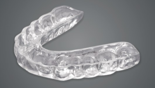
If your jaw clicks or pops when you open — or occasionally catches and momentarily locks before releasing — that's a different problem from the muscle-driven clenching a standard night guard is designed for, and it calls for a different kind of appliance. An anterior repositioning splint (ARS) is a removable acrylic appliance, most often worn on the upper teeth, built with an angled ramp that guides your lower jaw slightly forward when you bite into it. That forward position is the whole point of the design: it's meant to address a jaw joint disc that has slipped out of its normal spot, not to protect teeth from grinding.
What's Actually Happening in the Joint
Your jaw joint (the TMJ) has a small cartilage disc that normally sits between the ball of the jaw and the skull, cushioning the joint as it moves. In disc displacement with reduction — the condition an ARS is built for — that disc has shifted forward out of position but slips ("reduces") back into place when you open wide, which is what produces the clicking or popping sound. An ARS holds the jaw in a forward posture specifically to bring the condyle back into a normal relationship with that displaced disc.
Researchers describe two overlapping explanations for why this works. One theory holds that the forward position directly recaptures the disc, restoring a normal disc-condyle relationship that is then very gradually "walked back" toward a more natural position through controlled, incremental adjustment of the splint over time. A second, related theory is that the forward posture simply gives the disc room to slip back into place on its own while the tissues around the joint adapt and remodel. Either way, imaging studies have shown the therapy can also improve how stress is distributed across the joint and promote favorable remodeling of the jaw bone itself, alongside relieving both joint pain and the muscle tension that often comes with it.
Who an ARS Is Actually For
The primary candidate is someone with disc displacement with reduction causing pain, clicking, or occasional brief locking — this population also carries a meaningful risk (estimated in the range of 14–30%) of that clicking progressing to a more restrictive locked state if left unaddressed, which is part of why treating it isn't purely about the noise. An ARS is also sometimes used, often alongside physical therapy or a manual reduction technique performed by the dentist, for select cases of acute disc displacement without reduction — particularly in adolescents, where protecting the jaw joint's ongoing growth is an added consideration. A third scenario is disc displacement occurring alongside a retruded lower jaw or Class II bite relationship, where advancing the jaw forward can address the disc position and the bite at the same time. What an ARS is not for is routine nighttime clenching or grinding without joint involvement — that's a stabilization splint's job, not this appliance's.
What the Timing of a Click Actually Tells Us
Not every click means the same thing, and the timing matters diagnostically. In disc displacement with reduction, the classic finding is a reciprocal click: one click as you open your mouth, when the jaw's condyle catches and recaptures the displaced disc, and a second, usually softer click near the end of closing, when the disc slips back off the condyle into its displaced resting position. A click confined to just one of those two phases doesn't rule out the diagnosis — current diagnostic criteria also recognize a single-phase click paired with a click during side-to-side or forward jaw movement as meeting the pattern — but it does change what we're looking for and how we interpret it.
The timing within each phase carries its own information. An opening click that occurs early, right as you start to open, generally reflects milder displacement, since the disc is close enough to its normal position that the condyle catches it almost immediately. A late opening click — one that doesn't occur until you're opening wide — suggests more significant displacement, because the condyle has to travel further before it manages to recapture the disc. The gap between where the opening click occurs and where the closing click occurs (sometimes called the inter-click interval) is itself a rough gauge of how severe the displacement is: a wider gap generally points to a less favorable, harder-to-reduce disc position.
Perhaps the most clinically important pattern is a change over time. If a joint that has clicked for months or years suddenly stops clicking and, around the same time, you notice your mouth doesn't open as far as it used to, that combination is a signal the disc may no longer be reducing at all — a progression from disc displacement with reduction to a more restrictive displacement without reduction, sometimes described as a "closed lock." This kind of progression happens in an estimated 14 to 30% of DDwR cases, which is exactly why we don't dismiss a change in your jaw's clicking pattern as unimportant just because clicking itself is common and often benign. We also distinguish true disc clicks from crepitus (a grinding or crunching sound tied to joint surface changes rather than the disc) and from noise originating in the opposite joint, which is part of why a hands-on exam palpating each joint separately during opening and closing is part of an accurate diagnosis.
None of this means every click needs treatment. Painless reciprocal clicking is frequently a stable, lifelong finding that doesn't progress or require intervention — the research consistently shows this is the more common trajectory. What shifts our attention toward active management is pain that occurs at the moment of the click, tenderness directly over the joint, a click that has recently changed or disappeared, or a history of the jaw briefly catching or locking. Those features, not the mere presence of a click, are what point toward an ARS or another active treatment rather than simply monitoring.
Click or Crepitus? Two Very Different Sounds
Not every jaw noise is a click, and telling the two apart matters because they point to different problems. A click is short and sharp — a single, well-defined "pop" with a clear beginning and end, produced by the disc catching on or slipping off the condyle. Crepitus is a completely different sound: a continuous, coarse grinding or grating noise, often described as "bone-on-bone" or "gravel underfoot," that runs throughout the movement rather than occurring as a discrete event. Where a click points toward disc displacement, crepitus is the clinical hallmark of degenerative joint disease — wear and breakdown of the joint's bony surfaces, essentially an arthritic change within the TMJ.
Crepitus itself isn't uniform. Fine crepitus is subtle, sometimes so quiet it's only detected with a stethoscope during examination, while coarse crepitus is loud enough to be both felt and heard from several inches away and reflects more advanced bony change. The two sounds can also appear in sequence as a joint's condition evolves: a joint may start out clicking as a reducing disc, progress to locking as the disc stops reducing, and in some cases eventually develop crepitus as the joint surfaces themselves begin to break down from the altered mechanics. That said, this progression isn't universal or inevitable, and plenty of clicking joints never develop crepitus at all.
It's worth being upfront that sound alone is an imperfect diagnostic tool. Studies looking at how reliably clinicians can distinguish these sounds by ear and palpation alone have found real limitations — relying on the clinical exam by itself to diagnose degenerative joint disease has been shown to catch only a little over half of true cases. That's exactly why, when we suspect crepitus and degenerative changes rather than straightforward disc displacement, we don't rely on listening alone: imaging such as CBCT is what actually confirms bony changes like osteophytes, surface erosion, or joint-space narrowing. Interestingly, research has found the two don't map perfectly onto imaging findings either — in one study, clicking correlated with certain bony changes on imaging more strongly than crepitus did, a reminder that the sound your joint makes is a useful clue, not a standalone diagnosis. The practical takeaway for you is simple: a sharp, momentary snap points us toward evaluating for disc displacement and appliances like an ARS, while a sustained grinding sound shifts the conversation toward degenerative joint disease and, if needed, imaging to confirm it.
How We Formally Diagnose Degenerative Joint Disease
Because crepitus alone is such an imperfect diagnostic signal, the field's current diagnostic standard — the DC/TMD (Diagnostic Criteria for Temporomandibular Disorders) protocol — doesn't rely on listening by itself. A clinical diagnosis of degenerative joint disease (DJD) requires two things together: a patient-reported history of TMJ noise during jaw movement within the past 30 days, and crepitus actually detected by the examiner on palpation during that same exam. Either finding alone isn't enough; the criteria specifically require both the history and the physical exam finding to agree before DJD is called clinically.
Even with both criteria met, the clinical exam by itself is a blunt instrument. Validation studies of the DC/TMD protocol have found that history plus palpation alone catches only a little over half of true DJD cases when checked against imaging, with sensitivity in the range of roughly 55–60% and specificity that, while higher, still leaves real room for both false negatives and false positives. That's precisely why a clinical impression of DJD is treated as provisional rather than final — it tells us to look further, not that we've confirmed the diagnosis.
Confirmation is an imaging question. CT or CBCT imaging is considered the reference standard for verifying actual bony change in the joint, and the criteria distinguish clearly confirmatory findings — osteophytes, surface erosion, generalized sclerosis, or subchondral cysts — from findings classified as merely "indeterminate," such as isolated flattening of the condyle or a subtle increase in cortical density, which can appear as normal anatomic variation and don't by themselves confirm degenerative disease. This distinction matters clinically: an indeterminate finding on a scan is a reason for continued monitoring, not automatically a diagnosis of DJD.
Within a confirmed diagnosis, the field further separates osteoarthritis from osteoarthrosis — a distinction that isn't just semantic. Osteoarthritis refers to degenerative joint change accompanied by pain and inflammation, while osteoarthrosis describes the same underlying structural, bony changes occurring without pain, often identified incidentally on imaging taken for another reason. Two joints can look identical on a CT scan and receive very different treatment plans depending on which of these labels actually fits — because it's the presence of pain and active inflammation, not the bony change itself, that drives whether we intervene or simply monitor. This is also part of why a clicking joint and a crepitant joint aren't always managed the same way in practice: the joint's mechanical sound is only one piece of a diagnostic picture that also depends on your symptom history and, when indicated, targeted imaging.
How Treatment Differs for Osteoarthrosis vs. Osteoarthritis
Because osteoarthritis and osteoarthrosis share the same underlying degenerative process and differ only in whether pain is present, that single distinction is what actually drives how aggressively we treat the joint — not the amount of bony change visible on a scan. A painless, well-functioning degenerative joint (osteoarthrosis) is managed very differently from one causing pain and limited opening (osteoarthritis), even when the imaging looks similar.
When crepitus or bony change is present but the joint is painless and functioning normally, the emphasis stays conservative: reassurance that painless degenerative change is often a stable, adaptive process rather than something actively worsening; joint unloading through a soft diet, jaw rest, and controlling clenching or grinding habits that load the joint; and, when nighttime parafunction is a factor, a stabilization splint to protect the joint from that added strain. Analgesics, injections, and surgery generally aren't indicated for a joint that isn't causing symptoms — the plan is to monitor for any onset of pain, decreasing jaw opening, or progressive imaging changes, which would shift management toward the next tier.
Painful degenerative disease (osteoarthritis) follows a staged, escalating approach that starts conservative and reserves invasive options for cases that don't respond. First-line care combines the same unloading and behavioral measures with splint therapy and NSAIDs — the two approaches with the best evidence for improving both symptoms and the joint's imaging trajectory over time. Movement-based and behavioral therapies add meaningful benefit at this stage too: in the largest network meta-analysis of chronic TMD pain treatments to date, cognitive behavioral therapy (particularly when paired with biofeedback or relaxation training), therapist-guided jaw mobilization, and manual trigger-point therapy all showed moderate-to-high certainty evidence for pain relief, and supervised jaw exercises improved physical function as well — a reminder that encouraging movement and coping skills tends to outperform passive rest alone.
For pain or restricted opening that doesn't respond adequately to conservative care — especially if imaging shows the degeneration progressing — arthrocentesis (a minor procedure that flushes and lubricates the joint) becomes the next step, and pairing it with an injected adjuvant such as hyaluronic acid, a corticosteroid, or platelet-rich plasma has been shown to meaningfully outperform arthrocentesis alone, with pooled data showing a substantial additional reduction in pain scores along with improved jaw opening. Earlier intervention at this stage tends to carry a better prognosis than waiting. Surgical options — arthroscopy, arthroplasty, or joint replacement — are reserved for severe structural derangement or degeneration that hasn't responded to everything above; for the overwhelming majority of patients, the joint never needs to reach that point.
The practical takeaway is that a degenerative diagnosis on its own doesn't tell you what to do next. An asymptomatic, well-functioning joint with visible wear on a scan is watched and protected, not treated aggressively, while pain and functional limitation — regardless of how mild or severe the imaging looks — are what move a joint up this ladder from reassurance, toward splints and medication, and only toward procedures when conservative measures have genuinely been given a fair trial.
Where NSAIDs Fit In
Since NSAIDs come up as the first-line medication step on that ladder, it's worth understanding what the evidence actually supports and doesn't. Oral NSAIDs are the recommended first-line pharmacologic option for acute joint-related TMD pain, and they've been shown to reduce pain and improve how wide the jaw opens — but the evidence for any single drug, dose, or duration is still limited and generally low-quality, and NSAIDs work best as one piece of a broader conservative plan (jaw rest, soft diet, splint therapy, physical therapy) rather than as a stand-alone fix. A meaningful practical point: full anti-inflammatory benefit for joint pain can take up to about three weeks to become apparent, so a short trial of a few days isn't a fair test of whether an NSAID is helping.
Diclofenac is the single most-studied NSAID for this specific joint. In one open trial, a scheduled dosing regimen reduced TMJ arthritis pain by more than half over three months — a result comparable to splint therapy alone — though a shorter, placebo-controlled trial wasn't able to separate it from placebo, which fits the pattern of benefit building gradually rather than immediately. Naproxen has similarly reduced TMJ pain by more than half versus placebo in a double-blind trial, with a measurable difference from placebo only emerging around the three-week mark, and it's generally considered to carry a more favorable cardiovascular risk profile than some other NSAIDs. Ibuprofen is the common over-the-counter first choice for brief, acute flares and is effective for pain and opening, though in head-to-head TMD studies it hasn't consistently outperformed other conservative options.
A few prescribing principles carry across all of these: dosing on a scheduled, time-contingent basis (rather than only as pain strikes) is generally favored for joint inflammation, and the guiding rule is the lowest effective dose for the shortest duration that accomplishes the goal. NSAIDs aren't appropriate for everyone — they're generally avoided in active stomach ulcer disease or significant kidney disease, and used cautiously in cardiovascular disease, since long-term NSAID use carries some degree of cardiovascular and gastrointestinal risk that varies by which specific drug is used. Topical NSAID formulations applied directly over the joint are a reasonable option when someone wants to limit how much medication reaches the rest of the body. None of this is a substitute for a conversation with us or your physician about which option, if any, fits your specific health history — it's meant to give you a realistic sense of what this medication class can and can't be expected to do for a TMJ diagnosis.
Design and Wear Protocol
The classic design sits on the upper arch, though mandibular and digitally-designed, 3D-printed versions have shown comparable or, in some measures, better results for pain and jaw opening in recent research. There's no single standardized wear schedule across the field, but a commonly described approach starts with near-continuous wear — on for roughly the first three months except while eating or brushing — followed by a gradual transition to nighttime-only wear paired with incremental adjustment of the appliance, allowing the bite to resettle in a controlled, monitored way. Wearing an ARS full-time for an extended period without eventually tapering it carries a real risk of creating a lasting change to how your teeth meet.
What Treatment Looks Like, Step by Step
The process has a set order, and the first two steps decide whether the rest is worth doing. First, we guide your lower jaw forward until your front teeth meet edge to edge and the click disappears; that is the position the splint will hold. In studies, a splint built at this edge-to-edge position recaptured the disc on MRI in about 93% of cases, compared with about 55% when the jaw was held at the smallest forward position that still worked. Second, because we should not assume the disc has returned just because the click is gone, we confirm it with an MRI taken with that bite in place before the appliance is made.
The splint itself is a full-coverage acrylic appliance with small indentations and angled ramps that guide your jaw into the forward position, with even contact across the back teeth. You wear it around the clock, taking it out only to clean it and your teeth, for at least about three to six months, and we check in monthly. Then comes the part patients do not always expect: the “walk-back.” Once the joint is stable, we gradually trim the back of the splint, roughly a millimeter every four to six weeks, so your back teeth can settle into their natural bite while the disc stays put. At about six months we review the MRI and your bite and decide whether to keep trimming, switch to a soft splint, or use a step-back splint set slightly farther back. We consider treatment finished when your bite is stable and your jaw does not slide back after about six weeks without the splint.
Digital and 3D-Printed Splints
A splint can be made from a digital scan instead of impressions and plaster models, then 3D-printed or milled. Scanning removes several transfer steps where small errors creep in, lets us simulate how your teeth glide against the splint before it exists, and means a lost or damaged splint can be reprinted without starting over. The research so far is encouraging but small: digital splints perform as well as conventional ones, and printed and milled versions fit comparably. What matters most is still the same two things as above — the jaw position we record and the MRI that confirms it — not how the plastic is made.
What the Evidence Shows
A large network meta-analysis pooling 48 randomized trials across occlusal splint types found that ARS ranked highest of all splint designs for reducing pain specifically in joint-based (arthrogenous) TMD, and performed better than both no treatment and standard stabilization splints at resolving joint clicking. A Cochrane review reached a more measured conclusion, finding a small benefit over no treatment for clinically assessed joint pain in disc displacement with reduction at three and six months — though that finding rests on a single, fairly small study, which is a reminder that the evidence base, while positive, is still thinner than we'd like. In a head-to-head comparison against arthrocentesis (a minor joint-flushing procedure) for more complex presentations involving intermittent locking, a stabilization splint approach produced equivalent pain relief but resulted in better jaw opening — a useful data point when weighing a less invasive splint-based approach against a procedural one.
How Long Do the Results Last?
This is the question worth asking before you start. While the splint is in your mouth, results are strong: in one large group of patients, about 92% of joints had a normal disc position on MRI at the end of treatment. After the splint is stopped, that number drops. Twelve months later, about 73% still had a good result in that same group, and the share with an excellent result fell from roughly 65% to 43%. In another study, only about 41% of joints still showed a normal disc position six months after stopping. Older clinical studies that followed patients without MRI described similar slow slippage over several years.
The reassuring part is that your symptoms usually hold up better than the disc does. In one series, clicking fell from about nine in ten joints to about one in eight a year later, and pain improved even in joints where the disc had slipped forward again. Researchers think the tissue behind the disc adapts and takes over some of its cushioning job, so the joint can feel and work well without the disc sitting in a textbook position. It also helps to know that a standard stabilization splint relieves pain about as well; the repositioning splint is chosen when we specifically want to recapture the disc, not because it is a stronger painkiller. Older age, more advanced joint changes, longer-standing symptoms, and a disc that did not come back when the splint was first fitted all make a lasting result less likely, which is part of why we image first.
The Honest Limitations
Two tradeoffs are worth understanding before starting ARS therapy. First, the anatomic result doesn't always match the symptom result: in one MRI-based study following patients after treatment, only about 40.6% of joints had a normal disc-condyle relationship on imaging six months later, even though most patients' symptoms had improved and stayed improved. In practice, this means the splint often succeeds at calming pain and clicking without the disc necessarily staying in a textbook-normal position long-term — which is a reasonable outcome, but not the same as an anatomic "cure." Second, because the appliance works by holding the jaw somewhere other than its original bite position, coming off it can leave the back teeth not quite meeting the way they used to (a posterior open bite) or create a "dual bite" sensation if the taper is rushed. We manage that risk with a carefully paced weaning schedule and follow-up, and we talk through upfront that some patients need a period of bite adjustment, a bridging soft splint, or occasionally orthodontic or restorative work afterward to fully resolve it. None of this makes an ARS the wrong choice for a genuinely displaced disc — it just means it's a therapy that requires monitoring through its full course, not a device you're handed and left to manage on your own.
How This Differs From Your Standard Night Guard
It's worth being direct about this distinction because the two appliances are easy to conflate: a standard hard, full-coverage night guard is built to protect your teeth from grinding forces and sits in your normal bite position, while an ARS deliberately moves your jaw out of that normal position to address a structural problem inside the joint itself. Using the wrong one for your situation — for instance, wearing a plain night guard when the real issue is a displaced disc — won't address the clicking or catching, which is exactly why an accurate diagnosis of what's actually happening in the joint comes before we recommend either appliance.
Sources
- Predicting Anterior Repositioning Splint Efficacy in Disc Displacement With Reduction Using MRI-Based Texture and Quantitative Analysis: A Retrospective Study — Gao B, Dong Y, Shan Y, et al., Clinical Oral Investigations (2025)
- Physiological Effects of Anterior Repositioning Splint on Temporomandibular Joint Disc Displacement: A Quantitative Analysis — Chen HM, Liu MQ, Yap AU, Fu KY, Journal of Oral Rehabilitation (2017)
- Anterior Repositioning Splints in the Management of Temporomandibular Disc Displacement: A Network Meta-Analysis — Sakarya RE, Alawawda O, Akbulak UC, Journal of Oral Rehabilitation (2026)
- Temporomandibular Disorders — Ohrbach R, Sollecito T, Omolehinwa T, et al., in Burket's Oral Medicine, 13th ed. (2021)
- Comparative Efficacy of Digital 3D-Printed and Conventional Stable Occlusal Splints in the Treatment of Temporomandibular Disorders — Qin H, Liu Y, Miao H, et al., Journal of Oral Rehabilitation (2025)
- Digital Application of Three-Dimensional Diagnosis and Treatment With a Virtual Articulator — Lee KC, Park JH, Kim JC, et al., Journal of Esthetic and Restorative Dentistry (2024)
- Maxillary Occlusal Device Designed With an Anterior Platform for Guiding the Occlusal Adjustments During the Delivery Appointment: A Dental Protocol — Kois JC, Kois DE, Zeitler JM, Revilla-León M, Journal of Esthetic and Restorative Dentistry (2023)
- Pilot Clinical Comparison of Three Occlusal Splint Fabrication Techniques: A Preliminary Study — Torné-Durán S, Marco-Martínez L, Serrano-Belmonte I, Scientific Reports (2025)
- Step-Back Anterior Repositioning Splint Retraction for Temporomandibular Joint Disc Displacement With Reduction in Adult Patients — Li H, Shen D, Chen Z, Li J, Journal of Oral Rehabilitation (2023)
- Clinical Effect of Digitalized Designed and 3D-Printed Repositioning Splints in the Treatment of Anterior Displacement of Temporomandibular Joint Disc — Jin X, Chi W, BMC Musculoskeletal Disorders (2024)
- Can Anterior Repositioning Splint Effectively Treat Temporomandibular Joint Disc Displacement? — Ma Z, Xie Q, Yang C, et al., Scientific Reports (2019)
- Physical Therapy and Non-Surgical Manual Disc Reduction Combined With Anterior Repositioning Splint for Acute Disc Displacement Without Reduction of the Temporomandibular Joint in Adolescents — Fang Z, Yao Y, Fan S, et al., Clinical Oral Investigations (2024)
- A Novel Combination of Treatments for Acute Anterior Disc Displacement Without Reduction of Temporomandibular Joint — Song ZQ, Li CX, Gong ZC, Scientific Reports (2025)
- Effectiveness of Occlusal Splint Therapy in the Management of Temporomandibular Disorders: Network Meta-Analysis of Randomized Controlled Trials — Al-Moraissi EA, Farea R, Qasem KA, et al., International Journal of Oral and Maxillofacial Surgery (2020)
- Occlusal Interventions for Managing Temporomandibular Disorders — Singh BP, Singh N, Jayaraman S, et al., Cochrane Database of Systematic Reviews (2024)
- Is There a Superiority Between Arthrocentesis and Stabilizing Occlusal Splint for the Treatment of Anterior Disc Displacement With Reduction and Intermittent Block and Anterior Disc Displacement Without Reduction in TMJ? Randomized Clinical Trial — Corrêa-Silva M, de Carvalho TMI, Zambon CE, Peres MPSM, Machado GG, Oral Surgery, Oral Medicine, Oral Pathology and Oral Radiology (2024)
- The Efficiency of Anterior Repositioning Splints in the Management of Pain Related to Temporomandibular Joint Disc Displacement With Reduction — Pihut M, Gorecka M, Ceranowicz P, Wieckiewicz M, Pain Research & Management (2017)
- Diagnostic Value of Ultrasonography for the Detection of Disc Displacements in the Temporomandibular Joint: A Systematic Review and Meta-Analysis — Su N, van Wijk AJ, Visscher CM, Lobbezoo F, van der Heijden GJMG, Clinical Oral Investigations (2018)
- Temporomandibular Disorders — Scrivani SJ, Keith DA, Kaban LB, The New England Journal of Medicine (2008)
- Clinical Variables Associated With the Presence of Articular Pain in Patients With Temporomandibular Joint Clicking — Poluha RL, De la Torre Canales G, Bonjardim LR, Conti PCR, Clinical Oral Investigations (2021)
- Classification of Temporomandibular Joint Internal Derangement Based on Magnetic Resonance Imaging and Clinical Findings of 435 Patients Contributing to a Nonsurgical Treatment Protocol — Hegab AF, Al Hameed HI, Karam KS, Scientific Reports (2021)
- Correlation Between Direction and Severity of Temporomandibular Joint Disc Displacement and Reduction Ability During Mouth Opening — Litko M, Berger M, Szkutnik J, Różyło-Kalinowska I, Journal of Oral Rehabilitation (2017)
- Exploring the Association Between Clinical Features and CBCT Findings in TMJ Degenerative Joint Disease — Wu M, Lai H, Almeida FT, Friesen R, Journal of Oral Rehabilitation (2025)
- The Reproducibility of Temporomandibular Joint Vibrations Over Time in the Human — Zhang J, Whittle T, Wang L, Murray GM, Journal of Oral Rehabilitation (2014)
- Association of Radiographic and Clinical Findings in Patients With Temporomandibular Joints Osseous Alteration — Arayasantiparb R, Mitrirattanakul S, Kunasarapun P, et al., Clinical Oral Investigations (2020)
- Sound Analysis of Temporomandibular Joint Internal Derangements With Phonographic Recordings — Ogütcen-Toller M, The Journal of Prosthetic Dentistry (2003)
- Quantitative Description of Temporomandibular Joint Sounds: Defining Clicking, Popping, Egg Shell Crackling and Footsteps on Gravel — Leader JK, Boston JR, Rudy TE, et al., Journal of Oral Rehabilitation (2001)
- Artificial Intelligence-Enhanced Diagnosis of Degenerative Joint Disease Using Temporomandibular Joint Panoramic Radiography and Joint Noise Data — Choi E, Shin S, Lee K, et al., Scientific Reports (2025)
- Reliability and Validity of Axis I of the Research Diagnostic Criteria for Temporomandibular Disorders (RDC/TMD) With Proposed Revisions — Look JO, Schiffman EL, Truelove EL, Ahmad M, Journal of Oral Rehabilitation (2010)
- Functional, Physical and Psychosocial Impact of Degenerative Temporomandibular Joint Disease — Yap AU, Zhang XH, Cao Y, Fu KY, Journal of Oral Rehabilitation (2022)
- Validity of the Brief Diagnostic Criteria for Temporomandibular Disorders — Sharma S, Drangsholt M, Durham J, Alstergren P, Ohrbach R, Journal of Dental Research (2026)
- Prevalence of Degenerative Alterations of Osseous Temporomandibular Joint Structures in Cone Beam Computed Tomograms in a Consecutive Sample of Young Adults — Schnabl D, Pamminger D, Rohde S, et al., Clinical Oral Investigations (2025)
- Diagnosis of TMJ Degenerative Diseases by Panoramic Radiography: Is It Possible? A Systematic Review and Meta-Analysis — Schröder AGD, Gonçalves FM, Germiniani JDS, et al., Clinical Oral Investigations (2023)
- Pain Profiling of Patients With Temporomandibular Joint Arthralgia and Osteoarthritis Diagnosed With Different Imaging Techniques — Kothari SF, Baad-Hansen L, Hansen LB, et al., The Journal of Headache and Pain (2016)
- Interventions for the Management of Temporomandibular Joint Osteoarthritis — de Souza RF, Lovato da Silva CH, Nasser M, Fedorowicz Z, Al-Muharraqi MA, The Cochrane Database of Systematic Reviews (2012)
- Next Steps in Development of the Diagnostic Criteria for Temporomandibular Disorders (DC/TMD): Recommendations From the International RDC/TMD Consortium Network Workshop — Michelotti A, Alstergren P, Goulet JP, et al., Journal of Oral Rehabilitation (2016)
- Effectiveness of Treatments for Temporomandibular Joint Osteoarthritis: A Systematic Review and Meta-Analysis — Spadon-Brito LG, Souza CSP, Mélo AM, et al., Clinical Oral Investigations (2025)
- Management of Chronic Pain Secondary to Temporomandibular Disorders: A Systematic Review and Network Meta-Analysis of Randomised Trials — Yao L, Sadeghirad B, Li M, et al., BMJ (2023)
- Pharmacological Interventions for Painful Persistent Temporomandibular Disorders (TMD) — Beecroft EV, Penlington C, Allison JR, et al., Cochrane Database of Systematic Reviews (2021)
- Long-Term Changes of Temporomandibular Joint Osteoarthritis on Computed Tomography — Song H, Lee JY, Huh KH, Park JW, Scientific Reports (2020)
- Outcomes of Modified Arthrocentesis Using Concentric Needle and Cannula Technique With Sequential Viscosupplementation and Orthobiologics in Both TMJ Compartments — Januzzi E, Pauletto P, Cunha TCA, et al., Scientific Reports (2025)
- Reported Concepts for the Treatment Modalities and Pain Management of Temporomandibular Disorders — Wieckiewicz M, Boening K, Wiland P, Shiau YY, Paradowska-Stolarz A, The Journal of Headache and Pain (2015)
- Efficacy of Hyaluronic Acid in Temporomandibular Disorders Evaluated With Diagnostic Criteria for Temporomandibular Disorders (DC/TMD) — Kukreja P, Kukreja BJ, Marrapodi MM, et al., Journal of Oral Rehabilitation (2025)
- Pharmacological Management of Orofacial Pain — Romero-Reyes M, Arman S, Teruel A, et al., Drugs (2023)
- Pathophysiology of TMD Pain — Basic Mechanisms and Their Implications for Pharmacotherapy — Cairns BE, Journal of Oral Rehabilitation (2010)
- Orally Administered NSAIDs — General Characteristics and Usage in the Treatment of Temporomandibular Joint Osteoarthritis: A Narrative Review — Derwich M, Mitus-Kenig M, Pawlowska E, Pharmaceuticals (2021)
- Pharmacological Treatments of Temporomandibular Disorders: A Systematic Review Including a Network Meta-Analysis — Christidis N, Al-Moraissi EA, Barjandi G, et al., Drugs (2024)
- Assessing the Efficacy and Safety of Different Nonsteroidal Anti-Inflammatory Drugs in the Treatment of Osteoarthritis: A Systematic Review and Network Meta-Analysis Based on RCT Trials — JiaoYi P, YongQi S, KeChun G, et al., PLoS ONE (2024)
Related to This Article
Have a Dental Question of Your Own?
Reach out and we'll help you find the right next step.
Schedule Your Visit Call 212-877-7475