Early Cavities: Can We Just Watch Them? What "Class 2" on an X-Ray Means
Not every spot on an X-ray needs a filling. Here is what a depth score like "class 2" means, when watching is safe, and when a filling is the right call...
By Dr. Boris Zusin · Published November 6, 2026
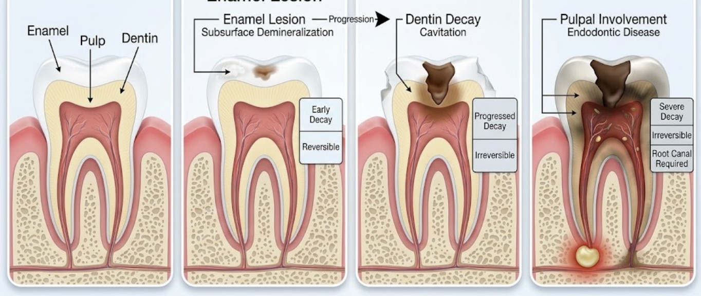
A spot on an X-ray is not always a cavity that needs drilling. Early lesions are caught while the tooth surface is still intact, and at that stage the goal is to stop them, not to fill them. Watching an early lesion is an active plan, not ignoring it.
What “Class 2” Means
Dentists score the depth of a shadow between teeth on a bitewing X-ray. A “2” (often called E2) usually means the shadow is in the inner half of the enamel and has not crossed into the softer layer underneath. That is an early lesion. Labels differ between offices, so it is fair to ask which scale was used.
Why Most Early Spots Are Not Holes Yet
When teeth are gently separated and the surface is examined directly, most early X-ray lesions turn out to be intact. In one study, about 9 in 10 inner-enamel lesions were still intact on the surface. They also tend to progress slowly, often over years. X-rays can also underestimate how deep a lesion is, which is why we track them over time.
What Watching Looks Like
- Fluoride: a stronger fluoride toothpaste at home and fluoride varnish in the office when needed.
- Cleaning where the spot is: flossing the contact and brushing the area carefully.
- Fewer sugary snacks and drinks between meals.
- Follow-up: exams every 3–6 months if your cavity risk is high, and X-rays at an interval set by your risk.
For active lesions in higher-risk patients, a thin sealant or resin infiltration can slow progression further without drilling.
Checking Whether the Surface Has Broken Down
If it is unclear, we may place a small elastic spacer between the teeth for a few days. This opens the gap slightly so we can look at the surface directly. It can be uncomfortable for a day or two, and it must be removed on schedule.
When We Recommend a Filling
- The surface has broken down and can’t be cleaned.
- The lesion reaches the middle layer of the tooth.
- It keeps growing despite good care.
- It causes symptoms.
Not sure what your X-ray shows? Schedule an exam and we will go through it with you.
Sources
- Dental Caries — Pitts NB, Zero DT, Marsh PD, et al., Nature Reviews Disease Primers (2017)
- When to Intervene in the Caries Process? An Expert Delphi Consensus Statement — Schwendicke F, Splieth C, Breschi L, et al., Clinical Oral Investigations (2019)
- Caries Management Pathways Preserve Dental Tissues and Promote Oral Health — Ismail AI, Tellez M, Pitts NB, et al., Community Dentistry and Oral Epidemiology (2013)
- Radiographic Assessment of Proximal Surface Carious Lesion Progression in Chilean Young Adults — Phillips M, Bernabé E, Mustakis A, Community Dentistry and Oral Epidemiology (2020)
- Detection of Cavitated Proximal Carious Lesions in Permanent Teeth: A Visual and Radiographic Assessment — Muñoz-Sandoval C, Gambetta-Tessini K, Botelho JN, Giacaman RA, Caries Research (2022)
- Temporary Tooth Separation to Improve Assessment of Approximal Caries Lesions: A School-Based Study — Nascimento MM, Ribeiro AP, Delgado AJ, et al., Operative Dentistry (2020)
- Sealing Proximal Non- and Micro-Cavitated Carious Lesions Using a One-Session Separator Technique: A 2-Year Randomised Clinical Study — Lindquist B, Emilson CG, Caries Research (2020)
Related to This Article
Have a Dental Question of Your Own?
Reach out and we'll help you find the right next step.
Schedule Your Visit Call 212-877-7475